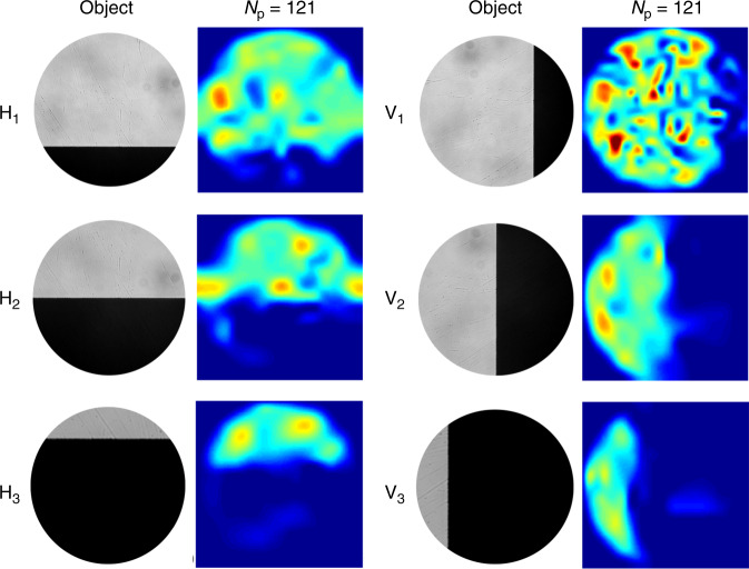
Fig. 2. COIL imaging of a knife edge using experimental data.
SARA–COIL results obtained using Np = 121 patterns. Micrographs of the objects are shown in the first and third columns, while corresponding SARA–COIL reconstructions are presented in the columns to the right of each set of objects. Hi and Vi respectively denote objects formed by horizontally and vertically overlaying a knife edge over ~25% (i = 1), ~50% (i = 2), and ~75% (i = 3) of the intensity pattern. Each reconstructed image has 125 × 125 pixels, with a field of view in the object plane of 0.9 mm × 0.9 mm.

