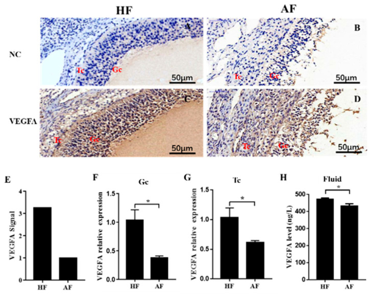
Figure 1.
Expression of VEGFA in healthy and atretic antral follicles. (A–D): Immunolocalization of VEGFA in healthy (A,C) and atretic (B,D) antral follicles; €: The signal intensity of VEGFA in follicles detected by the GeneChip Porcine Genome Array; (F,G): relative expression levels of VEGFA in GC and TC, respectively, detected by qRT-PCR; (H): expression level of VEGFA in the follicle fluid detected by ELISA. NC, negative control; HF, healthy follicle; AF, atretic follicle; GC, granulosa cell; Tc, theca cell; scale bar = 50 μm. Data are expressed as the mean ± SEM. * p < 0.05.

