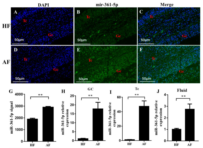
Figure 2.
Expression of the mir-361-5p in healthy and atretic antral follicles. (A–F): RNA-FISH was utilized to examine the localization of mir-361-5p in healthy and atretic antral follicles; (G): Signal intensity of mir-361-5p in follicles detected by µParaflo™ microfluidic chip; (H–J): relative expression levels of mir-361-5p in GC, TC, and follicle fluid, respectively, detected by qRT-PCR. HF, healthy follicle; AF, atretic follicle; GC, granulosa cell; TC, theca cell; scale bar = 50 μm. Data are expressed as the mean ± SEM. ** p < 0.01.

