Figure 1.
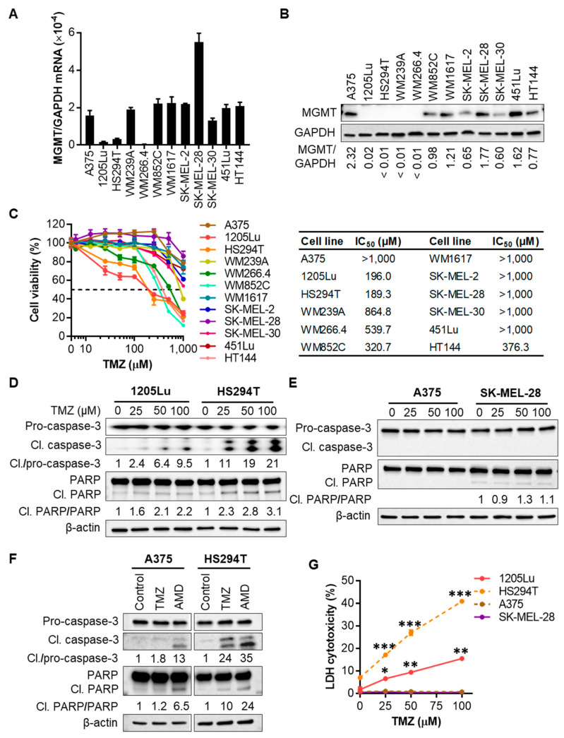
Melanoma cell lines with low expression levels of O6-methylguanine-DNA methyltransferase (MGMT) are sensitive to temozolomide (TMZ). (A) qRT-PCR analysis of MGMT mRNA expression in a panel of 12 human metastatic melanoma cell lines. (B) Western blot analysis of MGMT protein expression in 12 metastatic melanoma cell lines. The band densities of MGMT were digitally quantitated and normalized to those of the corresponding glyceraldehyde-3-phosphate dehydrogenase (GAPDH) in each cell line. (C) Melanoma cell viability evaluated by 3-(4,5-dimethylthiazol-2-yl)-5-(3-carboxymethoxyphenyl)-2-(4-sulfophenyl)-2H-tetrazolium (MTS) assay after the treatment with a single dose of TMZ at 0-1 mM for 72 h. Fifty percent inhibitory concentration (IC50) values were calculated using GraphPad Prism software. (D) Western blot analysis of caspase-3 cleavage and Poly(ADP-ribose) polymerase (PARP) cleavage indicated by the presence of cleaved (17, 19 kDa) caspase-3, and cleaved (89 kDa) PARP, respectively, in 1205Lu and HS294T cells treated with TMZ at 0–100 µM for 48 h. The band densities of cleaved caspase-3 and PARP were digitally quantitated and normalized in each cell line to those of the corresponding pro-forms subjected to the same treatment. (E) Western blot analysis of caspase-3 and PARP cleavages in A375 and SK-MEL-28 cells. (F) Western blot analysis of caspase-3 and PARP cleavages in A375 and HS294T cells treated with 100 µM TMZ for 48 h or apoptosis inducer actinomycin D (AMD; 10 µM) for 6 h. (G) Lactate dehydrogenase (LDH) release into culture medium measured using a colorimetric assay as a marker of cell death after TMZ treatment for 48 h. Data are representative of two independent experiments, and are expressed as the mean ± SEM (n = 3–4). * p < 0.05, ** p < 0.01, and *** p < 0.001 vs. the corresponding vehicle control.

