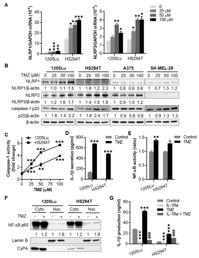
Figure 2.
Effects of TMZ on NACHT, LRR and PYD domains-containing protein (NLRP) expression and inflammasome activation. Where indicated, cells were treated with a single dose of TMZ for 48 h, then tested for (A) NLRP1 and NLRP3 mRNA expression by qRT-PCR, (B) NLRP1 and NLRP3 protein expression, as well as cleaved caspase-1 (p20 form) by Western blot, and (C) caspase-1 activity by fluorochrome-labeled inhibitors of caspases (FLICA) apoptosis assay. In (B), the band densities were quantitated and normalized to those of the corresponding loading control β-actin subjected to the same treatment. (D) Interleukin-1β (IL-1β) secretion analyzed by enzyme-linked immunosorbent assay (ELISA) from cells treated with 100 µM TMZ for 48 h. (E) Nuclear factor-κB (NF-κB) activity in cells treated with 100 μM TMZ for 48 h, determined using the Ready-to-Glow Secreted luciferase assay. (F) Western blot analysis of intracellular localization of NF-κB p65 in cells treated with 100 μM TMZ for 48 h. Cytoplasmic and nuclear fractions of cells were isolated and assayed for NF-κB p65 expression. Cyclophilin A (CyPA) and Lamin B were used as markers for cytoplasmic and nuclear proteins, respectively. The band densities of NF-κB p65 were quantitated and normalized to those of the corresponding loading control. (G) Intracellular IL-1β production analyzed by ELISA from cells treated with 100 µM TMZ and/or 10 µg/mL IL-1 receptor antagonist (IL-1Ra) for 48 h. Data are representative of two independent experiments, and are expressed as the mean ± SEM (n = 3). * p < 0.05, ** p < 0.01, and *** p < 0.001 vs the corresponding vehicle control. ### p < 0.001 vs the TMZ alone treatment.

