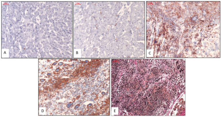
Figure 1.
Representative Histologic and Immunohistochemical Images. Images (A) & (B): Patients with normal myocardium; i.e., no CD3 stain (A) and no Mac1 stain (B). Images (C) & (D): Patients with IGCM presenting GCs surrounded by diffuse infiltration of massively increased T lymphocytes (CD3) (C) and macrophages (Mac1) in immunohistologic staining (D). Image (E): EvG staining from a patient with severe active myocarditis and giant cells. Magnification ×200.

