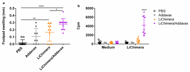
Figure 5.
Detection of cellular immune responses following LiChimera vaccination. The BALB/c mice were intramuscularly vaccinated two times at two-week intervals with LiChimera alone or adjuvanted with Addavax. (a) Delayed-type hypersensitivity (DTH) responses were evaluated on day 10 post boosting by measuring the difference between the thickness of the test and control footpads at 24 h post injection; (b) Spleens were collected two weeks post boosting and splenocytes were stimulated in vitro with LiChimera (2.5 µg/mL) for 72 h. Antigen-specific splenocyte proliferation was determined by [3H]-thymidine incorporation after another 18 h of culture and expressed as counts per minute (cpm). Data are expressed as means ± s.d. for n = 6 (PBS and Addavax) or n = 8 mice (LiChimera and LiChimera/Addavax). Significant differences between groups are indicated with asterisks measured by one-way ANOVA and Tukey’s multiple comparison tests. * p < 0.05, ** p < 0.01, and **** p < 0.0001.

