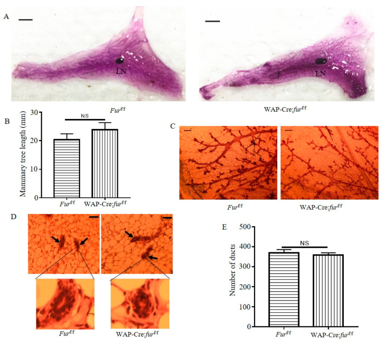
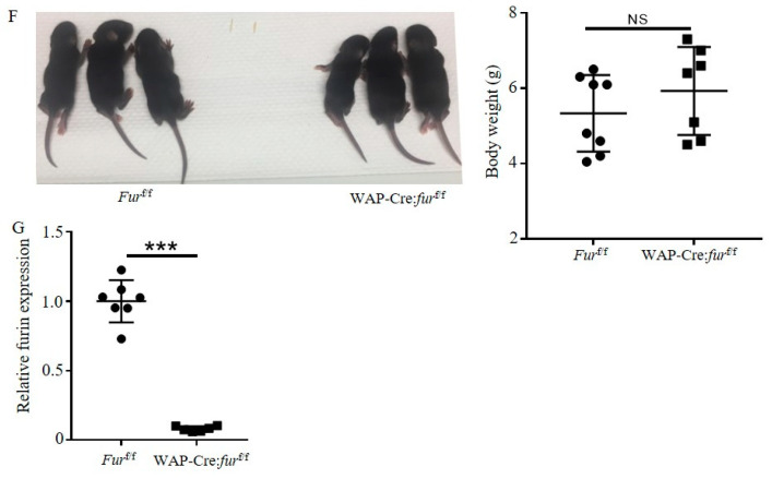
Figure 2.
Normal mammary development in mice with Furin deficiency in the mammary glands. (A) Representative photos of mammary gland whole-mount carmine alum staining for multiparous furf/f or WAP-Cre:furf/f female mice. Scale bar: 5 mm. LN: lymph node. (B) Quantification of length of the longest mammary tree past the lymph node in the whole mammary gland. n = 3 for each group. (C) Representative photos of whole-mount carmine alum staining of ductal epithelium from multiparous furf/f or WAP-Cre:furf/f female mice. Scale bar: 200 µm. (D) Representative H&E staining of mammary glands from multiparous furf/f or WAP-Cre:furf/f female mice. Scale bars: 50 μm. n = 3 for each group. (E) The number of ducts in H&E stained sections of mammary glands from multiparous furf/f or WAP-Cre:furf/f female mice. (F) The 10-day-old pups fed by a multiparous fur f/f female or WAP-Cre:furf/f female mouse (left panel). The body weight of 2 litters of 10-day-old pups fed by a multiparous furf/f female or WAP-Cre:furf/f female mouse (right panel). Each dot represents one pup. (G) The mRNA expression of Furin in mammary glands from multiparous furf/f female or WAP-Cre:furf/f female mice. A two-tailed Student’s t-test was performed for B, E, F and G. NS: not significant, *** p < 0.001.


