Figure 4.
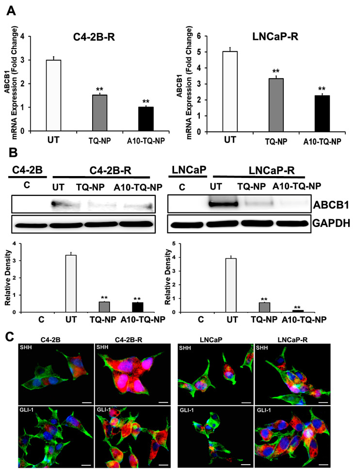
Gene expression changes in the ATP-binding cassette (ABC) transporter and the hedgehog (Hh) pathway in drug-resistant PCa cells. (A) Docetaxel (DTX)-resistant (C4-2B-R and LNCaP-R) PCa cells were treated with or without A10-conjugated TQ-NPs for 48 h. The expression levels of ABCB1 were measured by RT-PCR and expressed as the fold change relative to the controls (parental cells, C4-2B or LNCaP); 18S rRNA was used as an internal control for data normalization. The experiments were repeated thrice. The data, presented as means and standard errors of means (±SEM), were analyzed by Student’s t-test. The asterisks ** indicate p values ≤ 0.01. UT: untreated (DTX-resistant) (B) DTX-resistant PCa cells were treated with or without A10-conjugated TQ-NPs for 48 h, and ABCB1 protein expression was assessed by Western blots. GAPDH was used as an internal standard for equalizing the samples. The lower panel shows the densitometry of corresponding Western blots. UT: untreated (DTX-resistant); C: control (parent cells). The experiments were repeated thrice. The data are presented as means ± SEM; the asterisks ** indicate p values ≤ 0.01. (C) Immunofluorescence expression of SHH and GLI1 markers in PCa cells. The parental (C4-2B and LNCaP) and DTX-resistant (C4-2B-R and LNCaP-R) PCa cell lines were stained with anti-SHH and GLI-1 antibodies. Images were captured by an EVOS-FL fluorescent microscope with a 40× objective lens and with an appropriate filter. Red: SHH and GLI; green: F-actin cytoskeleton stained with Phalloidin 488; blue: nuclei counterstained with DAPI. Scale bar, 50 µM.

