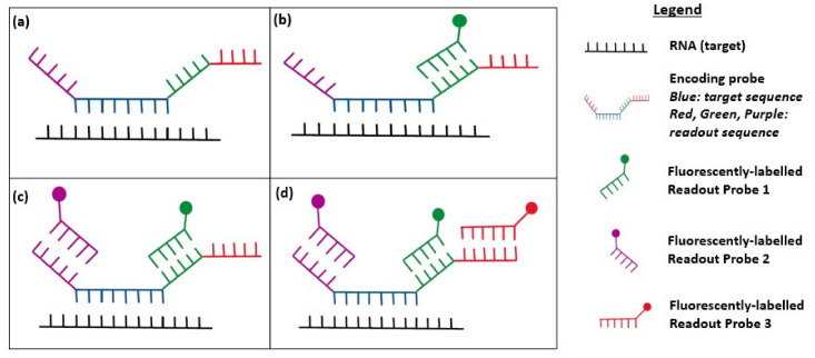
Figure 1.
Diagram showing multiplexed error-robust FISH (MERFISH) imaging. (a) Diagram depicting the encoding probe hybridized to the cellular RNA; (b) After the first round of hybridization, readout probe 1 (green) binds to the complementary readout sequence on the encoder probe and is fluorescently labelled (green circle); (c) After round 2 of hybridization, readout probe 2 (purple) binds to the encoding probe; (d) This process is repeated with N rounds of hybridization, whereby readout probe N (red) will bind to the readout sequence on the encoding probe.

