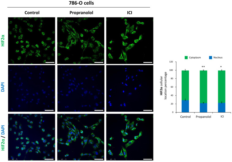
Figure 3.
Effect of β2-adrenergic receptor blockage on HIF-2α subcellular distribution. Left, immunofluorescence detection and confocal microscopy representative images from 786-O cells treated with 100 µM propranolol or ICI for 72 h. Mouse anti-human HIF-2α antibody stains (green), DAPI nuclear staining (blue) and merged are shown. Right, relative quantification of HIF-2α nuclei or cytoplasmic distribution. For quantification procedures, 12 different optic fields were taken from 4 replicates per condition. Scale bars represent 50 µm. Error bars denote ± SEM. Student’s t-test: * p < 0.05; ** p < 0.01; *** p < 0.001.

