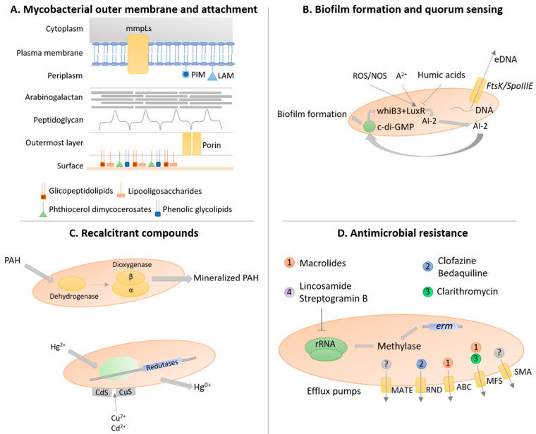
Figure 2.
The resilient biology of non-tuberculous mycobacteria. (A) Non-tuberculous mycobacteria (NTM) possess a unique outer membrane with several biolayers that increase cell hydrophobicity and environmental resistance. In particular, several classes of lipids are extremely important for mycobacteria sliding motility and cell attachment to the surface, triggering early biofilm formation. (B) Biofilm formation is quorum-sensing dependent with oxidative stress (ROS/NOS) and divalent cations (A2+) promoting whiB3 and luxR gene expression, with the consequent increase production of autoinducer-2 (AI-2), leading to the increase in cyclic diguanylate (c-di-GMP) activation and biofilm formation-associated gene expression. Contrary, humic acids inhibit gene expression of biofilm formation-related genes. Additionally, eDNA is secreted into the extracellular matrix by FtsK/SpoIIIE secretion system. (C) NTM are also resistant to recalcitrant compounds, such as polycyclic aromatic hydrocarbons (PAH) and heavy metals. The degradation of PAHs is achieved by a dioxygenase system composed of a dehydrogenase, the dioxygenase small (beta)-subunit, and the dioxygenase large (alpha)-subunit, while mercury (Hg2+) triggers the synthesis of mercuric reductases and copper (Cu2+) and cadmium (Cd2+) are chelated into the mycobacterial cell wall as sulfides. (D) NTM can resist antimicrobials by several mechanisms, including: the intrinsic cell wall provides a physical barrier towards the entrance of antimicrobials; the erm genes that cause the methylation of rRNA, resulting in resistance to macrolides, lincosamide, and streptogramin B; and the expression of efflux pumps of different superfamilies, namely multidrug and toxic compound extrusion (MATE), resistance-nodulation-cell division (RND), ATP-binding cassette (ABC), major facilitator (MFS), and small multidrug resistance (SMR), that actively secrete several antimicrobial compounds. The information present in this figure was combined from data reported by several studies regarding various non-tuberculous mycobacteria species.

