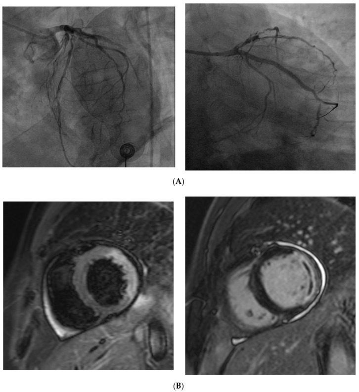
Figure 3.
(A) Coronary angiography of a 67 year old man presenting with STEMI shows the complete occlusion of a large marginal branch and a severe stenosis in the middle segment of the left anterior descending (left). Coronary angiography of the same patient demonstrates the revascularization of the marginal branch after percutaneous coronary intervention (PTCA) and drug-eluting stent (DES) implantation. (B) Cardiac magnetic resonance of the same patient, acquired the day after the PTCA shows alterations in the vascular territory of the previously occluded coronary vessel. Hyperintense signal on T2 turbo inversion recovery magnitude (TIRM) sequence reflects myocardial edema in the anterolateral and inferolateral segments (left). Delayed enhancement (hyperintense signal) indicates transmural infarction in the same myocardial segments (right); the linear hypointense area within the pathological myocardial hyperintensity, referred to as “no-reflow” phenomenon, is consistent with microvascular obstruction. It is due to a persistent perfusion defect despite the epicardial vessel revascularization, and is associated with a worse prognosis.

