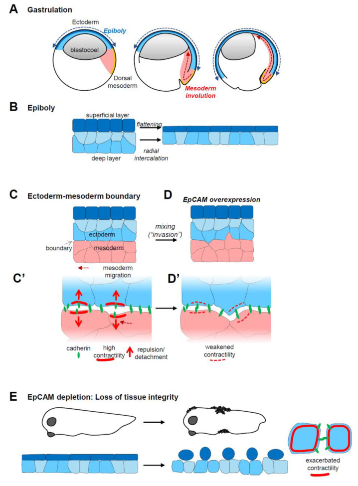
Figure 1.
EpCAM gain-of-function and loss-of-functionembryonic phenotypes. (A) Diagram of three consecutive stages of Xenopus gastrulation, indicating the movement of ectoderm epiboly (blue) and of mesoderm involution (red). (B) Epiboly involves two morphogenetic movements: The cells of the superficial layer flatten, while the cells of the deep layer rearrange by radial intercalation to rearrange into a single layer. The combined action of these two movements results in a large expansion of the surface of the ectoderm, which, at the end of gastrulation, covers the entire embryo. (C) The ectoderm and mesoderm are kept separated by a sharp interface, a so-called embryonic boundary. The mesoderm migrates along the surface of the ectoderm, using ectoderm cells as substrate for adhesion. (C’) The boundary results from ephrin-Eph-mediated repulsive reactions that locally boost actomyosin contractility, which leads to local and transient detachments of cadherin adhesions across the boundary. Through alternate attachments and detachments, mesoderm migration can proceed without intermingling with the ectoderm. (D) High EpCAM expression decreases actomyosin contractility, perturbing the function of the boundary. This results in mixing between the ectoderm and mesoderm layers, blocking mesoderm involution. (D’) At the cellular level, reduced contractility abolishes repulsive reactions, favouring intimate adhesive contacts between the two tissues, and eventually leading to their intermingling. (E) EpCAM depletion leads to massive loss of tissue integrity, due to uncontrolled contractility that results in cells’ rounding up and disaggregation.

