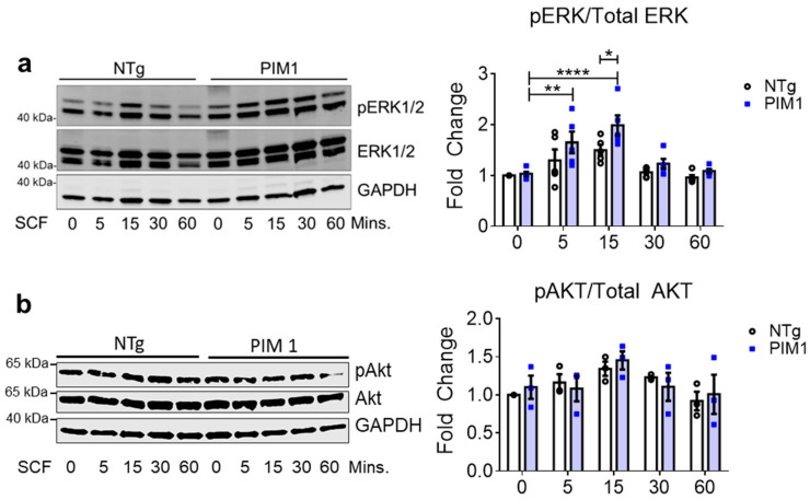
Figure 3.
Cardioprotective signaling downstream of c-Kit is elevated in PIM1 cardiomyocytes. (a) Immunoblot analysis of activated ERK1/2 and (b) activated AKT in NTg and PIM1 cardiomyocytes following treatment with SCF over 60 min. Quantification is shown on the right. N = 5, Error bars represent SEM, * p < 0.05, ** p < 0.01 and **** p < 0.0001 as measured by two-way ANOVA multiple comparison with Tukey.

