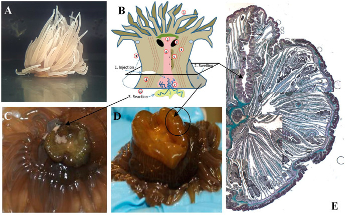
Figure 6.
Morphology variation after bacterial infection in A. viridis. (A), Schematic model of anatomy and injection site, swelling and reaction (B), Reaction zone (C), rejection and swelling of animal body 24 h after injection of suspensions of various heat-killed bacteria in (inset) the reaction after E. coli injection (D), A. viridis Gomori stain histological section (E), The original figure was produced for the study published by Trapani et al., [135]. The modified figure is consistent with the topic of the review.

