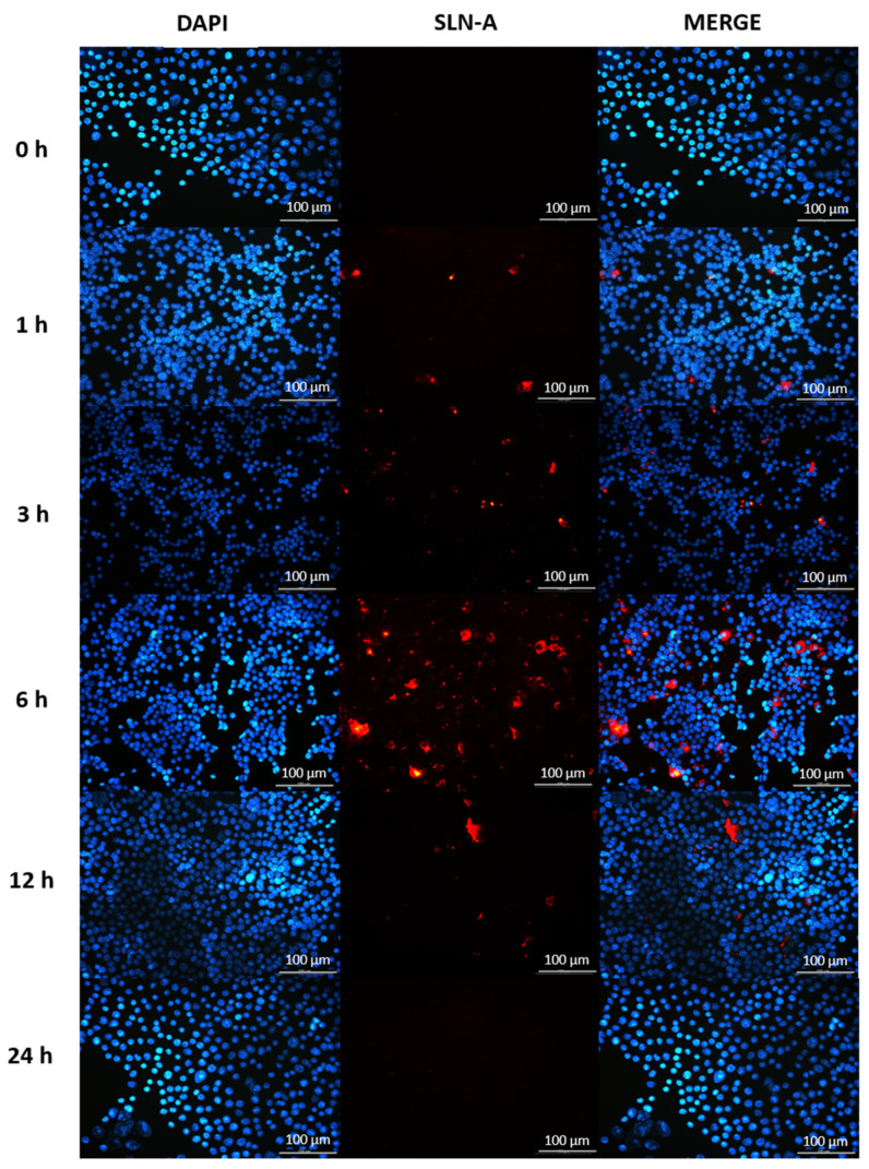
Figure 7.
Fluorescence micrographs of DC2.4 cells internalising SLN-A nanoparticles within 24 h. Cell nuclei counterstained with DAPI (blue) and SLN-A nanoparticles (red) are displayed. The speed of cell uptake of SLN-A nanoparticles by DC2.4 cells was visualised at 0, 1, 3, 6, 12 and 24 h timepoints. Particles were internalised from 3 h and were no longer visible within the cell by 24 h. Scale bar 100 µm.

