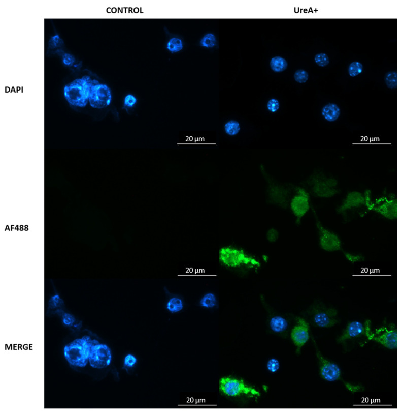
Figure 8.
Fluorescence micrograph of DAPI-stained DC2.4 cells (blue; nuclei) expressing Urease alpha (UreA) labelled with AlexaFluor 488 conjugated anti-His monoclonal antibody (green). Cells were transfected with lipoplex-A (SLN-A nanoparticles loaded with pcDNA3.1-UreA) and were found to be expressing UreA protein indicated by the binding of anti-6XHis antibody. Scale bar 20 µm.

