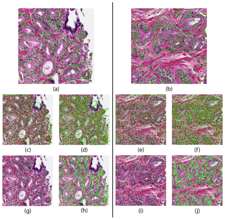
Figure 1.
Feature maps of a patient that did not progresses (left) and one who progressed (right). Nuclear segmentation results representing the nuclear shape (a,b). Nuclear spatial arrangement represented through the Delaunay triangulation (c,e) Voronoi tessellation (d,f), and Minimum Spanning Trees (g,i). Disorder in localized nuclear orientation (h,j).

