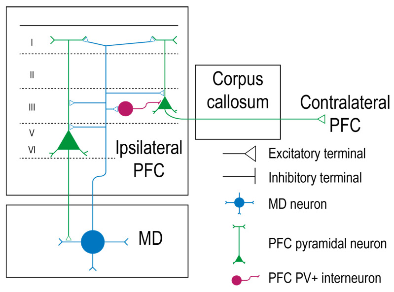
Figure 1.
Mediodorsal thalamic neurons have bidirectional connections with prefrontal cortex pyramidal neurons. The pyramidal neurons of the prefrontal cortex (PFC), represented in green, distribute their soma in layer 3, 5, and 6. The deep layer pyramidal neurons project back to the mediodorsal thalamus (MD), forming a loop. The mediodorsal thalamocortical neuron is represented in blue and synapses with the pyramidal neurons. The parvalbumine-expressing inhibitory interneurons (PFC-PV+) are represented in dark purple, while the excitatory connections are represented as a triangle [24].

