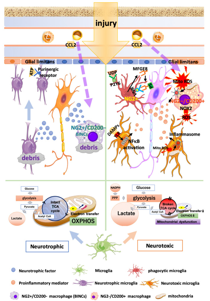
Figure 1.
Multiple functions of microglia and macrophages in brain injury. Neurotrophic microglia and macrophages (left half) exhibit an intact TCA cycle and stable mitochondrial OXPHOS. Microglia that have migrated to the injury site release anti-inflammatory cytokines and neurotrophic factors, thus encouraging wound healing and debris clearance. Neuroprotective infiltrated macrophages called BINCs (brain Iba1+/NG2+ cells) express a variety of neuroprotective factors. Neurotoxic microglia and macrophages (right half) produce energy in a glycolysis-dependent manner and exhibit increased lactate production, glucose uptake, and pentose phosphate pathway (PPP). DAMPs recognized by TLR stimulate NFκB pathways, leading to an increased expression of proinflammatory mediators. Microglia phagocytose viable neurons by recognizing opsonized PS via VNRs and the humoral “eat-me” signal UDP, through P2Y6. Phagocytic microglia and macrophages express the phagocytosis marker CD68 and NG2. Neurotoxic infiltrated macrophages (NG2−/CD200+ macrophages) release a greater amount of MitoROS, IL1β, and NOX2 compared with microglia. MitoROS not only directly damages the brain tissues, but also induces proinflammatory reactions by inducing the formation of inflammasomes.

