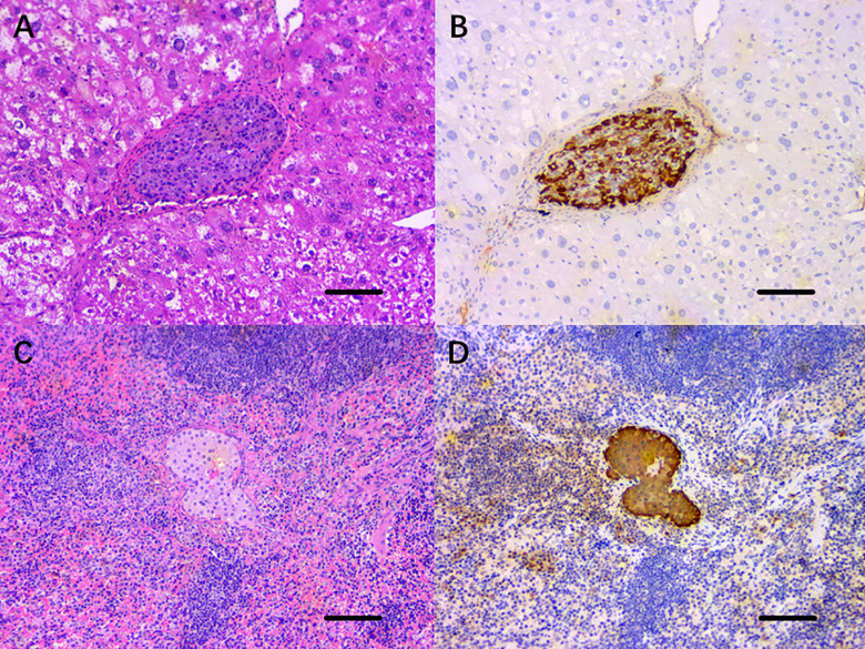
Fig. 6.
Histopathological staining of the islet grafts transplanted into the HST or the SP, 120 d posttransplantation. (A) H&E staining of the horizontal section of the left lobe of the liver revealed that the islet grafts in the HST were surrounded by normal liver cells and hepatic blood sinuses, with no obvious inflammatory cell infiltration. (C) H&E staining of horizontal section of the spleen showed that the islet graft was located in the splenic parenchyma, and no inflammatory cells infiltrated into the islet graft. (B, D) Immunohistochemical staining of the islet grafts sections for insulin in both groups showed that insulin was expressed uniformly in the cytoplasm of the islet cell mass. Scale bars, 100 μm.
H&E: hematoxylin and eosin; HST: hepatic sinus tract; SP: splenic parenchyma.

