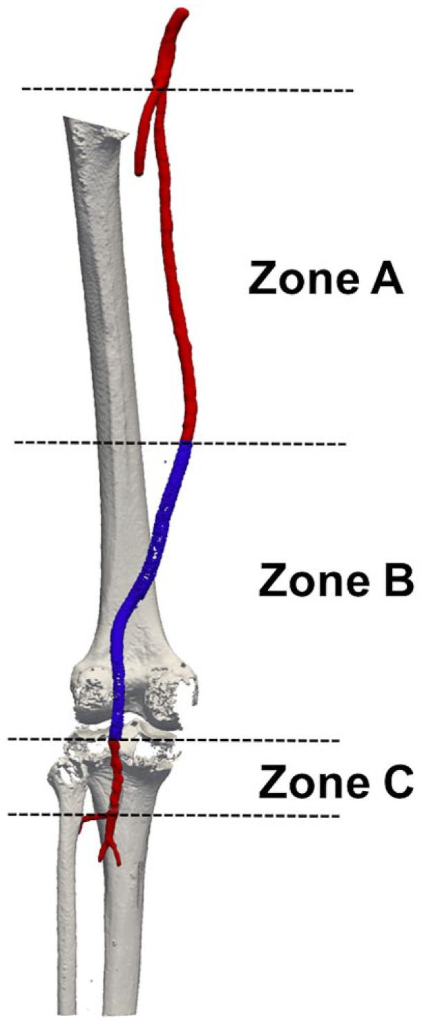
Figure 2.

Subdivision of the femoropopliteal segment into 3 zones: (A) between the origin of the superficial femoral artery (red) and the proximal end of the stent-graft (blue), (B) from the proximal to the distal end of the stent-graft (blue), and (C) from the distal end of the stent-graft to the origin of the anterior tibial artery (red).
