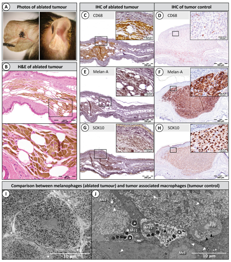
Figure 4.
Histopathology of ablated tumours (3 × 133.41 Gy). (A) A picture of the remaining nevus 18 months after MRT. (B) The nevus was formed by melanin-laden cells; (C,E,G) the ablated tumours stained immunohistochemically (IHC) and immediately bleached with trichloroisocyanuric acid (TCCA) to eliminate the excess of melanin; positive staining is shown with the 3,3′-Diaminobenzidine chromophore (DAB) as a bright yellow colour. Melanin-laden cells were positive for the macrophage marker CD68 (C), while they were negative for two melanoma markers: Melan-A (E) and SOX10 (G); (D,F,H) are positive controls in a melanoma harvested 10 days after implantation (not bleached); positive staining is shown with the NovaRED chromophore; (I) the ablated tumour under transmission electron microscopy revealed that the melanophages were packed with melanin and separated by dense collagen fibres (stars); (J) a group of macrophages (MΦ 1, 2, 3) in between melanoma cells (Me 1, 2, 3) 6 days after a single MRT. Macrophages contain numerous primary lysosomes (black arrowheads) and secondary lysosomes (asterisks). Melanin granules are indicated with white arrowheads.

