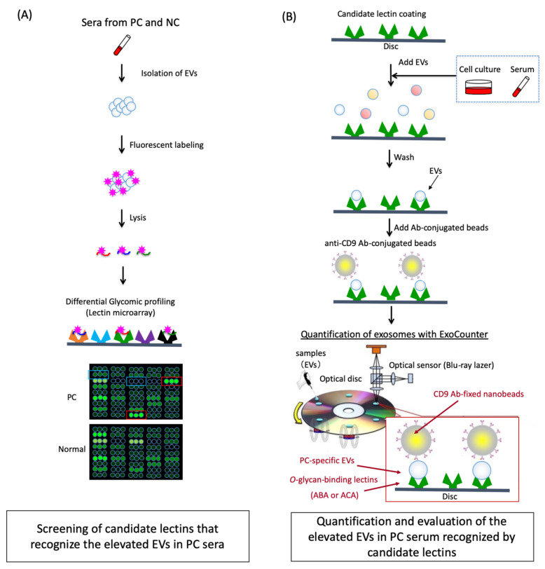
Figure 1.
Schematic strategy for screening and quantification of elevated extracellular vesicles (EVs) with specific glycans in pancreatic cancer (PC) sera. (A) Screening of candidate lectins that recognize the elevated EVs in PC sera. EVs in PC or normal control (NC) sera were isolated and labeled with fluorescent tags. The glycan profiles of the labeled EVs were then analyzed using lectin microarray, and the candidate lectins that recognize the elevated EVs in PC sera were identified by comparison with NC sera. (B) Quantification and evaluation of the elevated EVs in PC serum recognized by candidate lectins. The candidate lectins were coated on the optical disc of the ExoCounter system. The lectin-binding EVs in the PC sera or the cell lines were captured on the disc and labeled with the anti-CD9 Ab-conjugated nanobeads. The absolute numbers of labeled-EVs were quantified using the optical disc drive of the ExoCounter. NC, normal controls; PC, pancreatic cancer; EV, extracellular vesicles.

