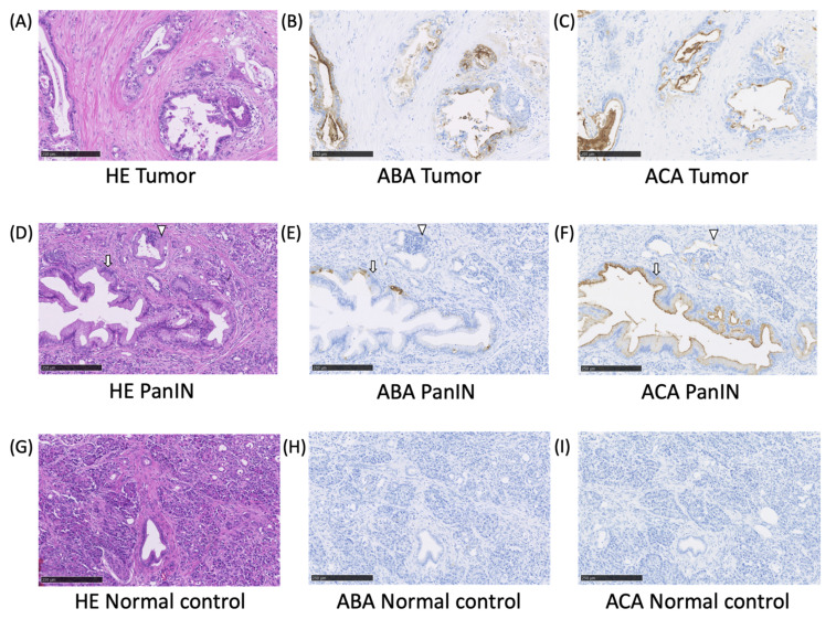
Figure 3.
Histochemical staining of PC and PanIN lesions using ABA and ACA. Formalin-fixed paraffin-embedded tissue sections from a PC patient were stained with the biotinylated ABA and ACA. The upper panel shows the invasive tumor (A–C), and the lower panel shows PanIN (D–F), and the normal pancreatic tissue (G–I). Hematoxylin-eosin: A, D, and G, ABA: B, E, and H, ACA: C, F, and I. PanIN and acinar-to-ductal metaplasia are indicated with an arrow and an arrowhead, respectively. The scale bars show 250 µm.

