Fig. 4.
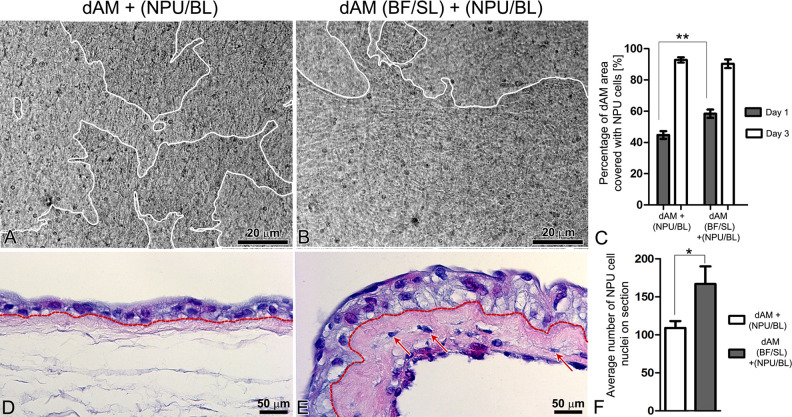
Analysis of normal porcine urothelial (NPU) cell growth and histological structure of the established urothelia. (A–C) After seeding for 24 h, the NPU cells cover a significantly larger area of the bladder fibroblast (BFs)-enriched de-epithelialized amniotic membrane (dAM) scaffolds (B) when compared with the dAM scaffolds alone (A); P < 0.001 (C). The white lines on A and B surround area of the dAM, overgrown with the NPU cells. On the third day of cultivation, the difference in the areas covered by the NPU cells, seeded on two different scaffolds is no longer significant; P > 0.05 (C). (D, E) After 3 weeks in culture, the NPU cells on the dAM scaffolds form two-layered to three-layered urothelium (D), whereas urothelia established on the BFs-enriched dAM scaffolds consist of 3–4 layers of NPU cells (E). Dashed lines indicate the AM basal lamina. (F) The significant difference in the stratification of the urothelia between the scaffolds is confirmed by counting the nuclei of the established urothelia; P < 0.05. (C) The graph presents the mean percentage of the dAM scaffolds covered with the NPU cells (N = 73 analyzed images of NPU cells on dAM scaffolds and N = 71 analyzed images of NPU cells on BFs-enriched dAM scaffolds, all from four independent experiments). (F) The average number of the NPU cell nuclei on the 7 µm thick and 2 mm long segments of the urothelium (N = 7 segments of the urothelium for dAM scaffold and N = 5 segments of the urothelium for BFs-enriched dAM scaffolds, from two independent experiments) ± SE * P ≤ 0.05, ** P < 0.001.

