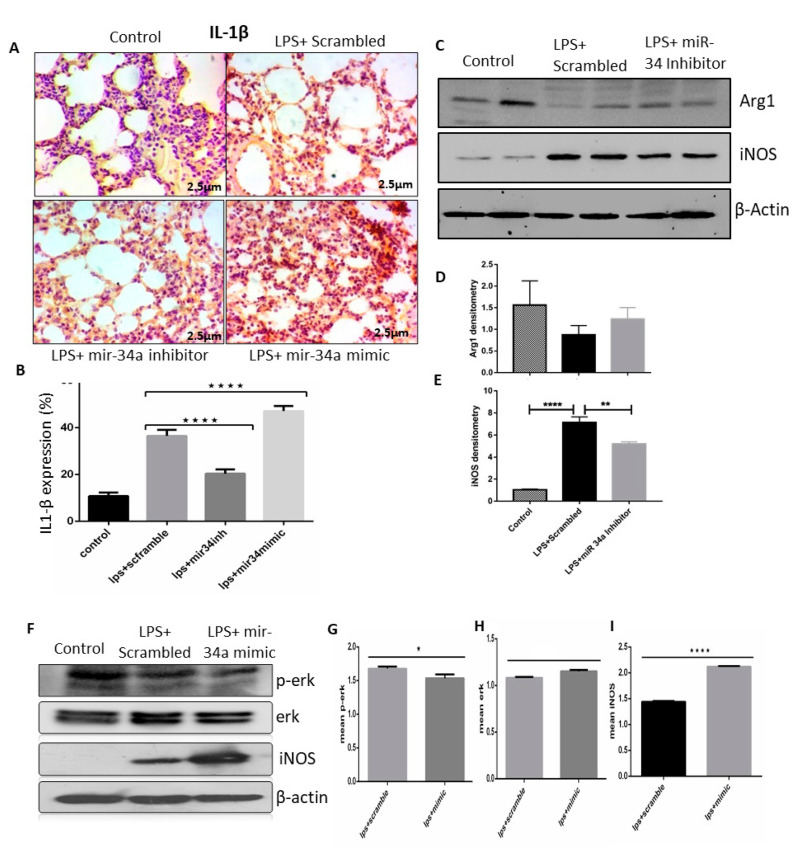
Figure 6.
LPS effectively induced miR-34a, which promoted the M1 phenotype of macrophages. (A) Lung IHC staining of control, LPS + scrambled, and miR-34a + LPS treated mice groups showed expression of IL1-β. (B) Color density of IL1-β was quantified and analyzed using image J and two-way ANOVA following Tukey’s post hoc analyses. (C–E) Immunoblots of Arg1 and iNOS were performed in lung tissue lysates of mentioned groups. Densitometry was performed for all immunoblots and analyzed by two-way ANOVA following Tukey’s post hoc analyses. (F–I) RAW macrophages transfected with miR-34a inhibitor or scrambled; after treatment with LPS, cell lysates were performed to check the expression of ERK, p-ERK, and iNOS in given groups. Densitometry was done, and t-tests were used for analysis. * p < 0.05, ** p < 0.01, **** p < 0.0001 (n = 4).

