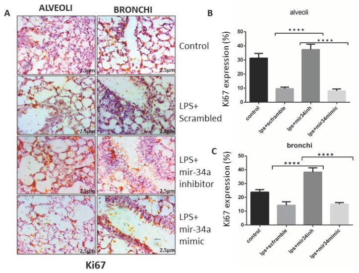
Figure 7.
miR-34a reduced cell proliferation. (A) Immune-histological images of mice lung tissue showed the expression of Ki67 after treatment with LPS along with miR-34a inhibitor, mimics, and scrambled treated groups. (B,C) Expression of Ki67 quantified in alveolar and bronchial areas and statistically analyzed by two-way ANOVA. **** p < 0.0001 (n = 4).

