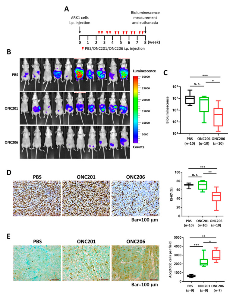
Figure 2.
Effect of ONC206 on uterine tumor growth and apoptosis in vivo. (A) Schema shows the use of an in vivo model to evaluate the effect of ONC206 on uterine tumor growth. i.p., intraperitoneal; PBS, phosphate-buffered saline. (B) Images show lower bioluminescence signals in ARK1 cell-bearing nude mice treated with ONC201 (n = 10) or ONC206 (n = 10) than in untreated PBS controls (n = 10). (C) Box plot shows a significantly lower bioluminescence from ARK1 cell-bearing nude mice treated with ONC206 (n = 10) than those treated with ONC201 (n = 10; p < 0.05, Mann–Whitney U test) or untreated PBS controls (n = 10; p < 0.001, Mann–Whitney U test). (D,E) Representative microscopic images of paraffinized sections of tumor tissues collected from ARK1 cell-bearing nude mice 6 weeks after treatment with ONC201 or ONC206, showing significantly (D) lower Ki-67 expression and (E) higher apoptosis in the ONC206 treatment group compared with the ONC201 treatment group or untreated controls. Bar = 100 μm. Quantification of cell staining positively for Ki-67 or apoptotic cells for each group is shown in the box plot. *** p < 0.001, ** p < 0.01, * p < 0.05, n. s. = not significant (p > 0.05), Mann-Whitney U test.

