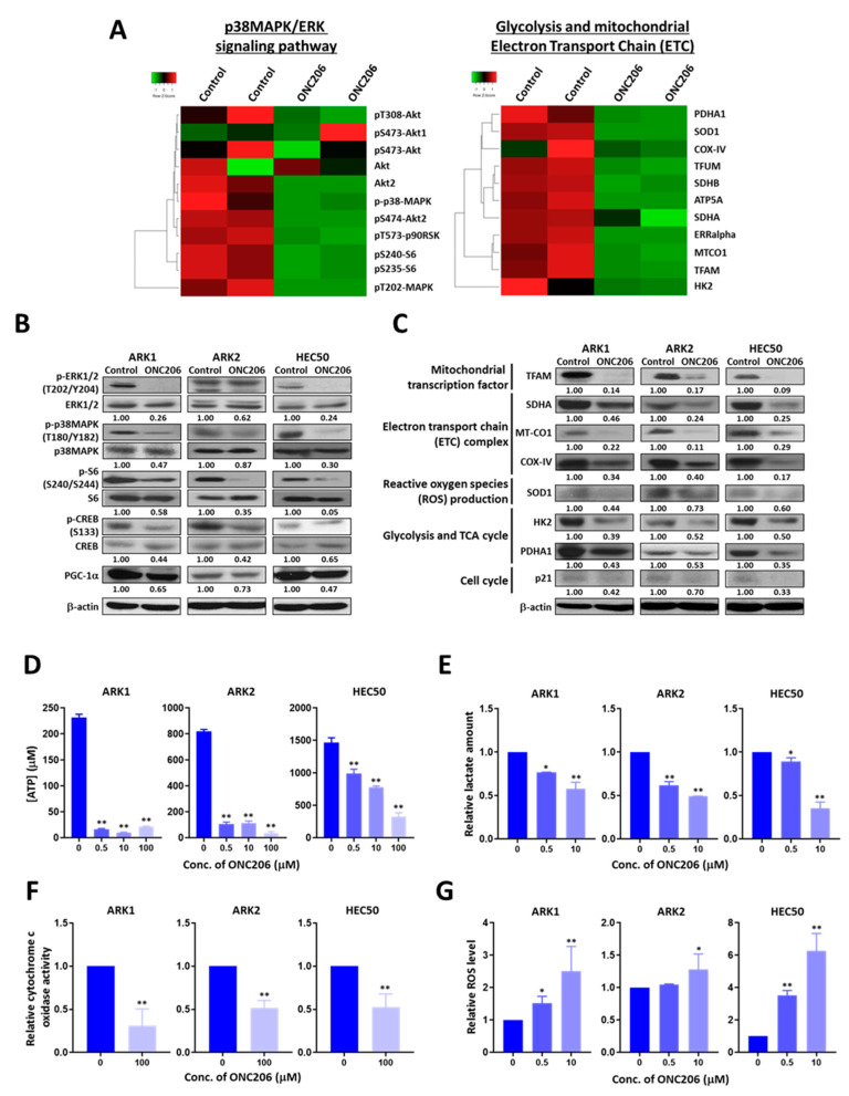
Figure 3.
Effect of ONC206 on mitochondrial functions and glycolysis in uterine serous cancer cells. (A) Heat maps obtained using reverse phase protein array analysis shows differentially expressed proteins related to the p38MAPK/ERK signaling network and mitochondrial functions in ARK1 cells with (n = 2) or without (n = 2) 48 h of treatment with 50 μM ONC206. (B) Western blot analyses show decreased protein levels of p-ERK1/2, p-p38MAPK, p-S6, p-CREB, and PGC-1α in ONC206-treated ARK1, ARK2 and HEC50 cells compared with control cells without treatment. β-actin served as a loading control. Ratios between phosphorylated and total protein levels are presented. Three independent experiments were performed. (C) Western blot analyses show lower protein levels of TFAM, SDHA, MT-CO1, COX-IV, SOD1, HK2, PDHA1, and p21 in ONC206-treated ARK1, ARK2, and HEC50 cells compared with control cells without treatment. β-actin served as a loading control. Relative normalized protein levels with respect to the corresponding control are presented. Three independent experiments were performed. (D) ATP production as measured by the ATP production kit, (E) lactate secretion as measured by the Lactate-Glo assay kit, and (F) cytochrome c oxidase activity as measured by the cytochrome c oxidase assay kit were decreased in ARK1, ARK2, and HEC50 cells after treatment with ONC206. Results were averaged from three independent experiments and are shown as mean ± standard deviation. ** p < 0.01, * p < 0.05, two-tailed Student t test. (G) Reactive oxygen species (ROS) productions was increased in ARK1, ARK2, and HEC50 cells after 24 h of treatment with ONC206 as measured using the ROS detection assay kit. Results were averaged from three independent experiments and are shown as mean ± standard deviation. ** p < 0.01, * p < 0.05, two-tailed Student t test.

