Figure 4.
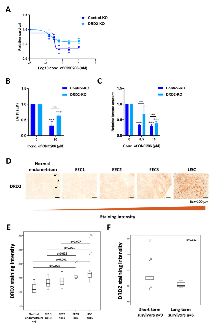
Role of DRD2 in mediating the effect of ONC206 on uterine serous cancer (USC) cells. (A) DRD2 knockout (DRD2-KO) ARK2 cells are more resistant to ONC206 than control knockout (Control-KO) ARK2 cells, as measured by the MTT assay. Three independent experiments were performed (mean ± standard deviation). (B) Decrease in ATP production in DRD2-KO ARK2 cells after 48 h of treatment with ONC206 as measured by the ATP determination kit. Results were averaged from three independent experiments and are shown as mean ± standard deviation. *** p < 0.001, ** p < 0.01, two-tailed Student t test. (C) Decrease in lactate secretion in DRD2-KO ARK2 cells after 24 h of treatment with ONC206 as measured by the Lactate-Glo kit. Results were averaged from three independent experiments and are shown as mean ± standard deviation. *** p < 0.001, ** p < 0.01, two-tailed Student t test. (D) Representative microscopic images of paraffinized sections of different stages of gynecological tissues including normal endometrium, endometrioid endometrial cancer (EEC; grades 1 to 3) and uterine serous cancer (USC). An increase in DRD2 expression from normal endometrium to USC is shown. Bar = 100 μm. Arrows indicate the uterine gland structures in normal endometrium. (E) Quantification of DRD2 staining intensity for normal endometrium (n = 5), grade 1 EEC (n = 10), grade 2 EEC (n = 10), grade 3 EEC (n = 5) and USC (n = 15) is shown in the box plot. (F) Box plot shows a significant decrease in DRD2 expression in long-term USC survivors (>5 years; n = 6) compared with short-term survivors (<2 years; n = 9; p = 0.012, Mann–Whitney U test).

