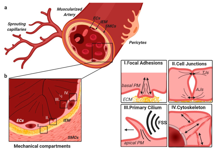
Figure 2.
Muscularized arteries are composed of different layers of cells and interconnective ECM (a); from inside to outside—ECs, the IEM, SMCs, and pericytes (b). In ECs, mechanical signaling is found at four major hubs (enlarged on the right): I. Focal adhesion-ECM contact sites, important in cellular migration, and adhesion. II. Cell–cell junctions that are highly important in the regulation of EC barrier function and cell–cell communication. This includes AJs, TJs, and gap junctions that contribute differently. III. The primary cilium, which is apically located in ECs and initiates signaling upon deflection by FSS, for example. IV. The cytoskeleton that links the plasma membrane with the nucleus, thereby allowing direct force transmission. Abbreviations: ECs—endothelial cells, IEM—inner elastic membrane, SMCs—smooth muscle cells, FSS—fluid shear stress, AJs—adherens junctions, TJs—tight junctions, and PM—plasma membrane.

