Figure 3.
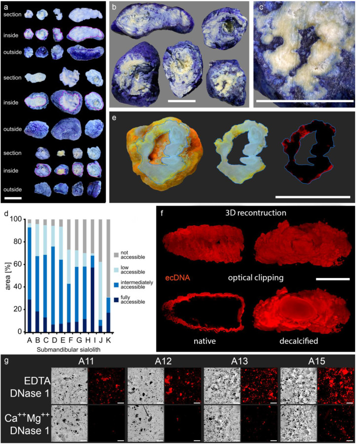
Sialoliths contain dye-impermeable, densely calcified areas and extracellular DNA (ecDNA). Stones were immersed in different dyes and subsequently ground to reveal their interior. The calcified surface is very compact and prevents the penetration of all employed dyes into the stones´ interior. (a–c) Trypan blue (MW = 873 g/mol) incompletely penetrates the native calculi, which is shown here in submandibular sialoliths (n = 11). (d) Morphometry of the submandibular sialoliths depicted in (a–c): every single stone shows dye inaccessible parts, particularly in the inner layers, quantified by image analysis (n = 11). (e) Acridin orange (MW = 265 g/mol) reveals that the surface of sialoliths occasionally communicates with cavities in the interior via small clefts (displayed in red, right panel; n = 5). (f) Representative LSFM (light sheet fluorescence microscopy) of a submandibular sialolith (n = 4). In native stones, propidium iodide (red, MW = 668 g/mol) only stains the extracellular DNA (ecDNA) at the surface. After decalcification, the dye penetrates the sialolith, demonstrating the role of the calcification for the compactness of the surface and indicates the containment of extracellular DNA within the entire stone, including the core. (g) Representative propidium iodide staining of stone gravel reveals the widespread distribution of extracellular DNA within the stones. The signals attenuated after treatment with DNase 1, confirming extracellular DNA as the signal´s origin (n = 7). Scale bar (a–c,e,f): 10 mm; scale bar (g): 200 µm.

