
Figure 2.
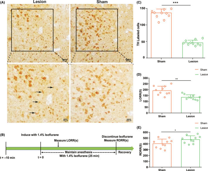
Effect of unilateral DA neuron lesion in the ventral periaqueductal gray (vPAG) on LORR and RORR time of isoflurane anesthesia. A, Representative immunohistochemical image showing unilateral site of lesion group and sham group in the vPAG. The lesion group animals were selectively depleted of DA Neurons in the vPAG with unilaterally injection of 1 μL of 6‐OHDA. Black arrowhead denotes the dead cells. B, Timeline for quantifying induction and recovery time with isoflurane. C, Quantitative analysis of the number of tyrosine hydroxylase‐positive neurons on the lesion and normal sides. D, Induction time and E, recovery time in the lesion and sham groups (n = 10 per group; mean ± SD; *P < .05; **P < .01)