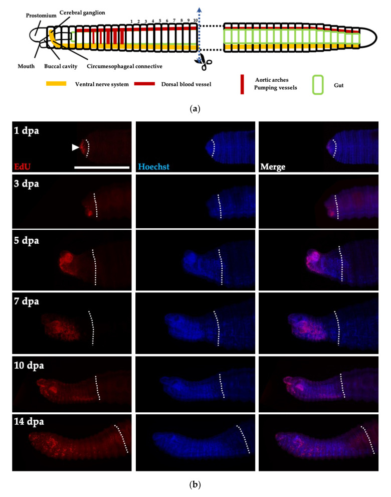
Figure 5.
Active cell proliferation in regeneration. (a) The schematic cartoon indicating the amputation site, which is located in the trunk region, lacking cell proliferation. (b) Cell proliferation in regenerating tissues is visualized by EdU staining (red) at indicated days post-amputation (dpa). Nuclei are marked by Hoechst staining (blue). Dotted lines indicate the site of amputation. Scale bar, 500 μm.

