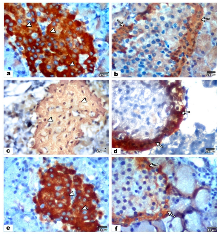
Figure 2.
Photomicrographs of immunohistochemically stained pancreatic sections. Control sections (a,b). (a) An anti-insulin immune-reactivity of pancreatic islet showing brown-stained pancreatic β cells with strong positive immune-reaction (arrowheads). (b) An anti-glucagon immune-reactivity of pancreatic islet showing brown-stained pancreatic α cells with mild positive immune-reaction (arrow). VPA treated pancreatic sections (c,d). (c) An anti-insulin immune-reactivity of pancreatic islet showing brown-stained pancreatic β cells with mild positive immune-reaction (arrowheads). (d) An anti-glucagon immune-reactivity of pancreatic islet showing brown-stained pancreatic α cells with strong positive immune-reaction (arrow). VPA and ALA treated pancreatic sections (e,f). (e) An anti-insulin immune-reactivity of pancreatic islet showing brown-stained pancreatic β cells with strong positive immune-reaction (arrowheads). (f) An anti-glucagon immune-reactivity of pancreatic islet showing brown-stained pancreatic α-cells with mild positive immune-reaction (arrow).

