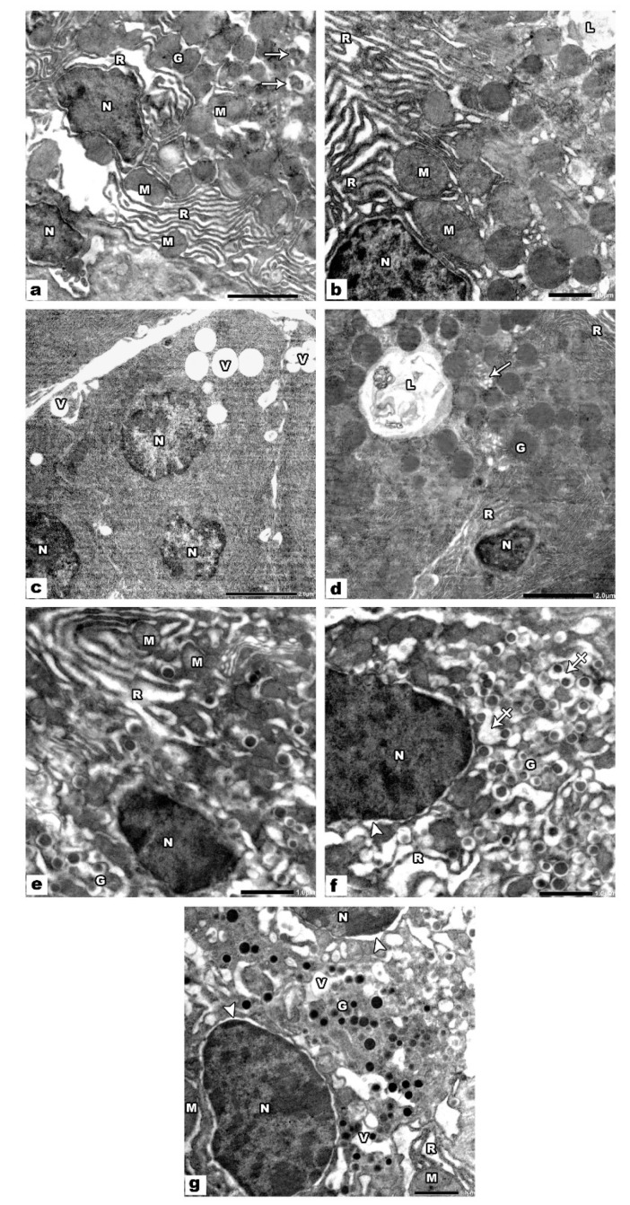
Figure 4.
Electron micrographs of VPA treated pancreatic sections. (a–d) The pancreatic acini show small dense irregular heterochromatic nuclei (N), dilated disordered rough endoplasmic reticulum (R), swollen mitochondria (M), decreased secretory granules (G) some of them appear degraded (arrows), many cytoplasmic vacuoles (V) and autophagic vacuoles (L). (e,f) β cells of the islets show small dense irregular heterochromatic nuclei (N), some nuclei (N) show perinuclear halo (arrowhead), dilated rough endoplasmic reticulum (R), swollen mitochondria (M), increased lucent halo of many of the secretory granules (G) with the fusion of some of them (crossed arrows). (g) α cells of the islets show small irregular condensed nuclei (N) with wide perinuclear space (arrowhead), vacuolated cytoplasm (V), dilated disordered rER (R), swollen mitochondria (M), and secretory granules (G).

