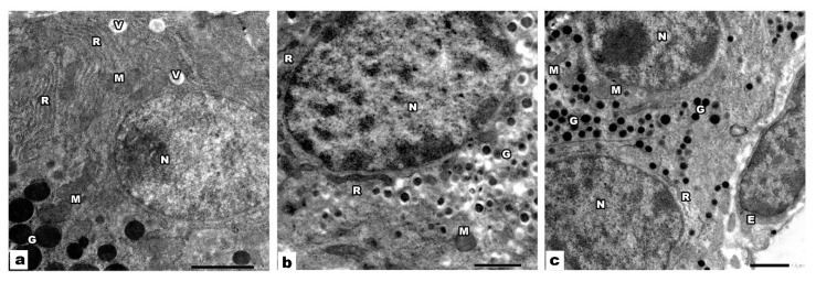
Figure 5.
Electron micrographs of VPA and ALA treated pancreatic sections. (a) The acinar cells have rounded euchromatic nuclei (N), the cytoplasm shows parallel cisternae of rER (R), mitochondria (M), electron-dense granules (G), and small vacuoles (V). (b) β cells of the islets of Langerhans have euchromatic nuclei (N), electron-dense granules encircled by halo (G), mitochondria (M), and rER (R). (c) α cells have euchromatic nuclei (N), many electron-dense granules (G), rER (R), and mitochondria (M). Note the endothelial cell (E) of the blood capillary.

