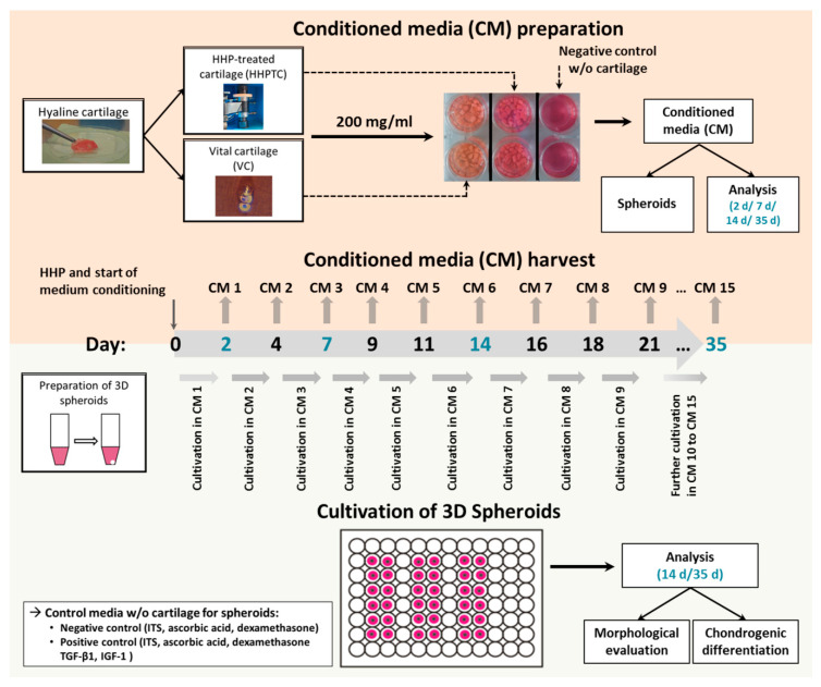
Figure 2.
Flow diagram of experimental approach and methods. The upper panel (light orange) shows the preparation of conditioned medium of HHP-treated and vital hyaline cartilage tissue. The tissue slices were cultivated for a total period of 35 days. Medium was changed and harvested every two to three days for subsequent use for 3D spheroid cultures. In addition, the release of soluble chondrogenic mediators was analyzed on day 2 (conditioned medium (CM 1), day 7 (CM 3), day 14 (CM 6) and day 35 (CM 15). The lower panel (light green) depicts the preparation and cultivation of chondrocytic spheroid cultures. After a two-day incubation in basal medium (negative control medium), spheroids were supplied with the respective CM of vital and HHP-treated cartilage. Spheroids, cultivated with basal medium containing ITS™, ascorbic acid and dexamethasone (negative control) or basal medium plus chondrogenic growth factors TGF-β1 and IGF-1 (positive control) served as controls. After 14 d and 35 d of incubation, spheroids were analyzed regarding morphological changes and chondrogenic differentiation.

