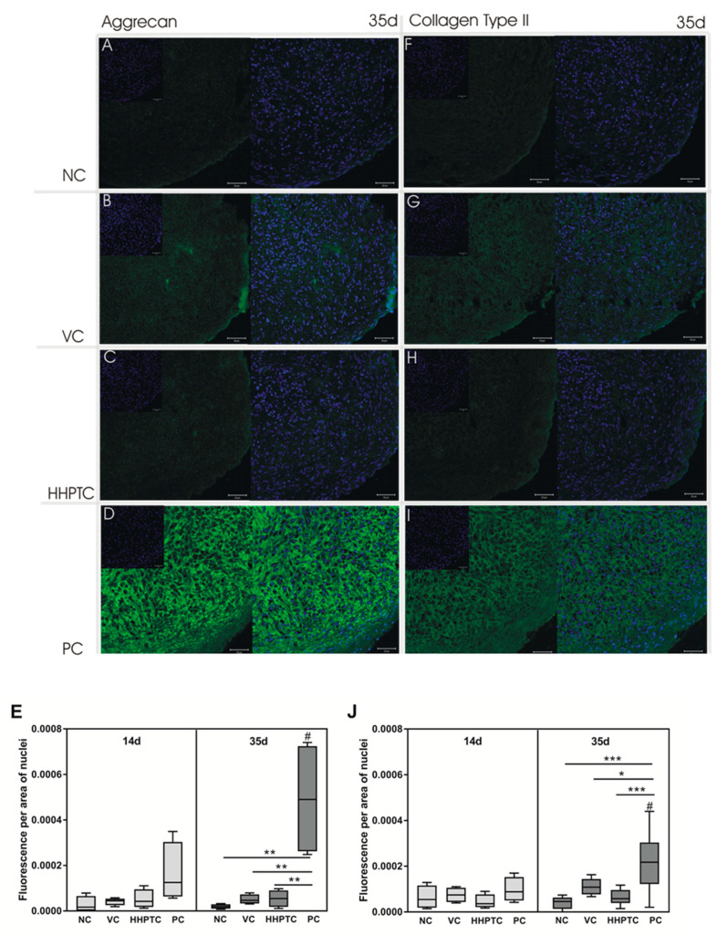
Figure 9.
Aggrecan and collagen type II protein expression. Immunohistochemical staining of chondrocytic spheroid cultures at days 14 and 35 was performed using anti-human aggrecan and anti-chicken collagen type II antibody (green fluorescence). Nuclei were stained with Hoechst (blue fluorescence). Chondrocytic spheroids were cultivated in following media groups: Negative control (NC) (A,F), vital cartilage conditioned medium (VC) (B,G), high hydrostatic pressure treated cartilage conditioned medium (HHPTC) (C,H) and positive control (PC) (D,I). Exemplary images from day 35 depict aggrecan (A–D) and collagen type II (F–I) staining. Magnification: 200×, scale bar: 50 µm. For semi-quantitative analysis (E,J), the relative fluorescence signal was determined with ImageJ. After background correction, mean fluorescence signal was related to area of nuclei (n = 8). Data are presented as boxplots with minimum, 25th percentile, median, 75th percentile and maximum. Statistical analysis was performed using Repeated Measures Two-way ANOVA with Bonferroni’s multiple comparison as post hoc test. Significance: * p < 0.05, ** p < 0.01, *** p < 0.001 between treatments and # p < 0.05 comparison between day 14 and 35.

