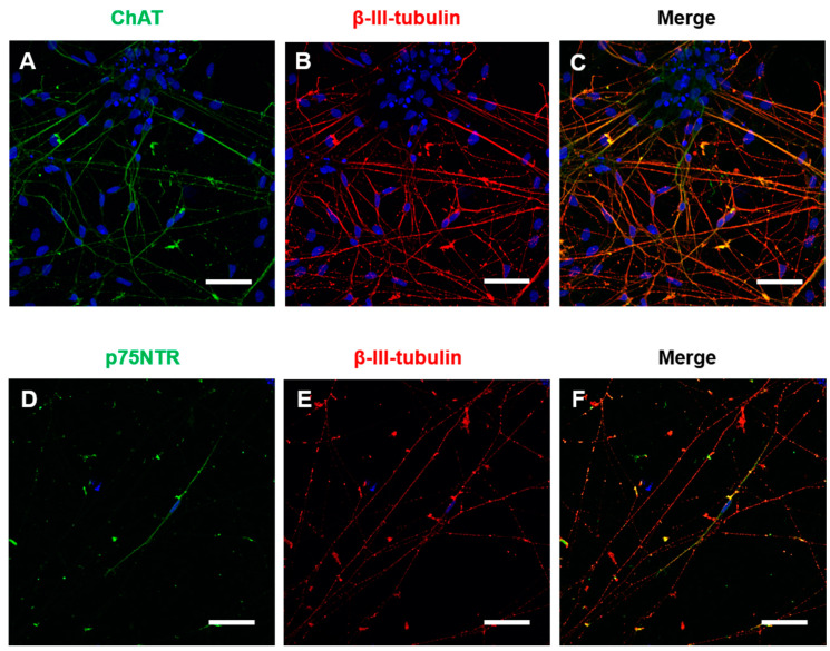
Figure 2.
BFCNs were positive for neuronal markers. BFCN cultures were matured for 4 weeks and immunocytochemistry for general neuronal markers and specific cholinergic neuronal markers was performed. (A–F) Representative confocal pictures of the BFCN cultures at week 4 of BFCN maturation with the nuclear marker Hoechst 33,342 in blue. (A) Cholinergic marker ChAT in green. (B) Neuronal marker β-III-tubulin in red. (C) Merged picture of A and B. (D) Cholinergic marker p75NTR in green. (E) Neuronal marker β-III-tubulin in red. (F) Merged picture of D and E. Scale bars = 50 μm.

