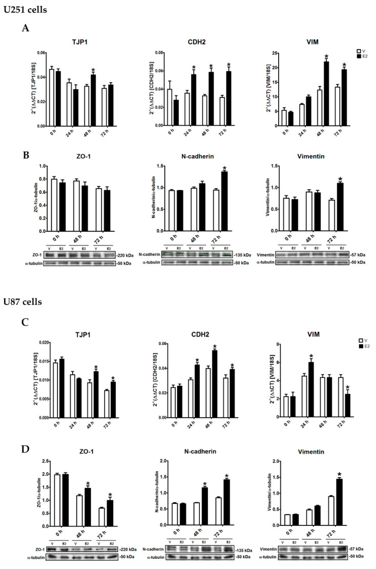
Figure 4.
E2-regulated epithelial-to-mesenchymal transition (EMT) marker expression of human GBM-derived cells. (A,B) U251 and (C,D) U87 cells were treated with 17β-estradiol (E2, 10 nM) and vehicle (V, 0.01% cyclodextrin) for 24, 48, and 72 h. (A,C) Epithelial gene (tight junction protein 1 (TJP1)) and mesenchymal genes (vimentin (VIM) and cadherin-2/N-cadherin (CDH2)) expression was quantified by RT-qPCR using the comparative method 2ΔΔCt concerning the reference gene 18S rRNA. (B,D) Zonula occludens 1 (ZO-1), N-cadherin, and vimentin content was determined by Western blot. Densitometric analysis of EMT marker expression with their respective representative bands using α-tubulin as a load control showed that E2 increased EMT marker expression with different temporal dynamics. Results are expressed as the mean ± standard error of the mean (SEM); n = 3; * p < 0.05 vs. V.

