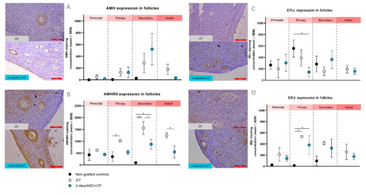
Figure 5.
(A) AMH, (B) AMHRII, (C) ERα, and (D) ERβ staining concentrations (mean ± SEM) comparisons between groups (non-grafted controls, OT, and 2-step/ASCs+OT), and follicle stages (primordial, primary, secondary, and antral) were analyzed using Kruskal–Wallis and Fisher’s post hoc LSD tests. Significant differences between groups are indicated as follows: * p < 0.05; *** p < 0.001. AMH: anti-Müllerian hormone. AMHRII: anti-Müllerian hormone receptor type II. ER: estrogen receptor. OT: ovarian tissue. ASCs: adipose tissue-derived stem cells. Scale bars: 100 µm.

