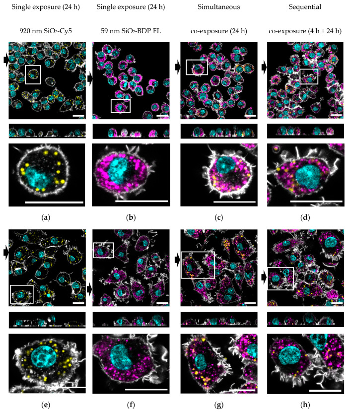
Figure 5.
Representative confocal laser scanning micrographs with the corresponding xz projections demonstrate the uptake of 59-nm SiO2-BDP FL NPs and 920-nm SiO2-Cy5 particles in J774A.1 macrophages. Upper panels (a–d): Cells exposed to single 920-nm SiO2-Cy5 particles (yellow), single 59-nm SiO2-BDP FL NPs (magenta), or a combination of SiO2 particles simultaneously for 24 h or sequentially for 4 h and 24 h. Bottom panels (e–h): Cells were prestimulated with 1-µg/mL LPS for 24 h and then post-exposed to single 920-nm SiO2-Cy5 particles, single 59-nm SiO2-BDP FL NPs, or a combination of both particles simultaneously for 24 h and sequentially for 4 h and 24 h. F-actin was stained as the cytoskeleton marker using a rhodamine-phalloidin conjugate (grey), and the nucleus was stained using 4′,6-diamidino-2-phenylindole (DAPI) (cyan). The black arrows represent the position of the xz projection (at the bottom), showing the intracellular localization of the particles. Zoom-in images (shown under each representative image) clearly demonstrate the cellular distribution of two types of SiO2 particles. Scale bar: 20 µM.

