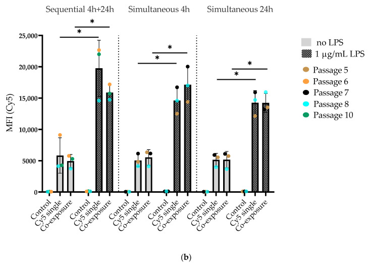
Figure 7.
Flow cytometry data showing the uptake of SiO2 particles in J774A.1 macrophages. Cells were prestimulated with 1-µg/mL LPS for 24 h and then exposed to a single particle type or a combination of 59-nm SiO2-BDP FL and 920-nm SiO2-Cy5 particles. For comparison, we used unstimulated cells exposed only to SiO2 particles in the same manner. The particles were administered sequentially or simultaneously for different time points (4 h and 24 h). (a) Median fluorescence intensity (MFI) of the BDP FL signal. (b) Median fluorescence intensity (MFI) of the Cy5 signal. Data show a significant increase (* p < 0.05) in the MFI of the BDP FL and Cy5 signals after LPS prestimulation. No significant difference in the MFI was observed between the single- and co-exposure conditions. Control cells were stained with DAPI and were not exposed to SiO2 particles.


