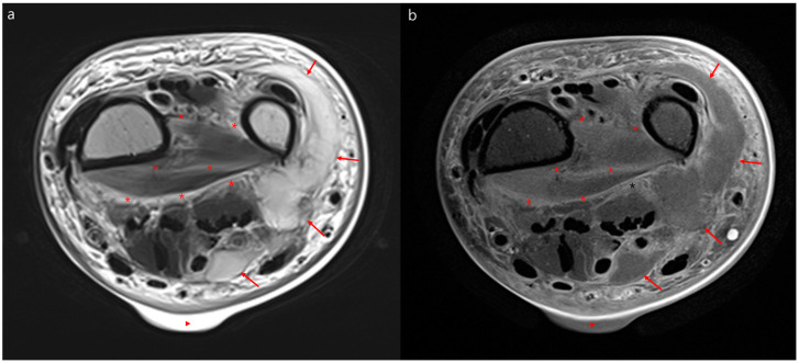
Figure 2.
Representative magnetic resonance images of necrotizing fasciitis. Necrotizing fasciitis of the left wrist in a 71-year-old woman. Axial T2 weighted magnetic resonance image (a) and contrast-enhanced magnetic resonance image (b) showing diffuse hyperintensity with irregular enhancement of the deep peripheral fascia and intermuscular deep fascia (asterisk) of the wrist. Additionally, there is a lobulating abscess in the ulnar side of the wrist (arrows) and a skin bulla (triangle).

