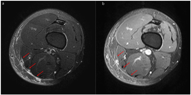
Figure 3.
Representative magnetic resonance images of severe cellulitis. Severe cellulitis of the left thigh in a 44-year-old man. (a) Fat-suppressed axial T2-weighted magnetic resonance image and a contrast-enhanced magnetic resonance image (b) showing localized hyperintensity within the deep peripheral fascia (arrows) with enhancement in the posteromedial thigh.

