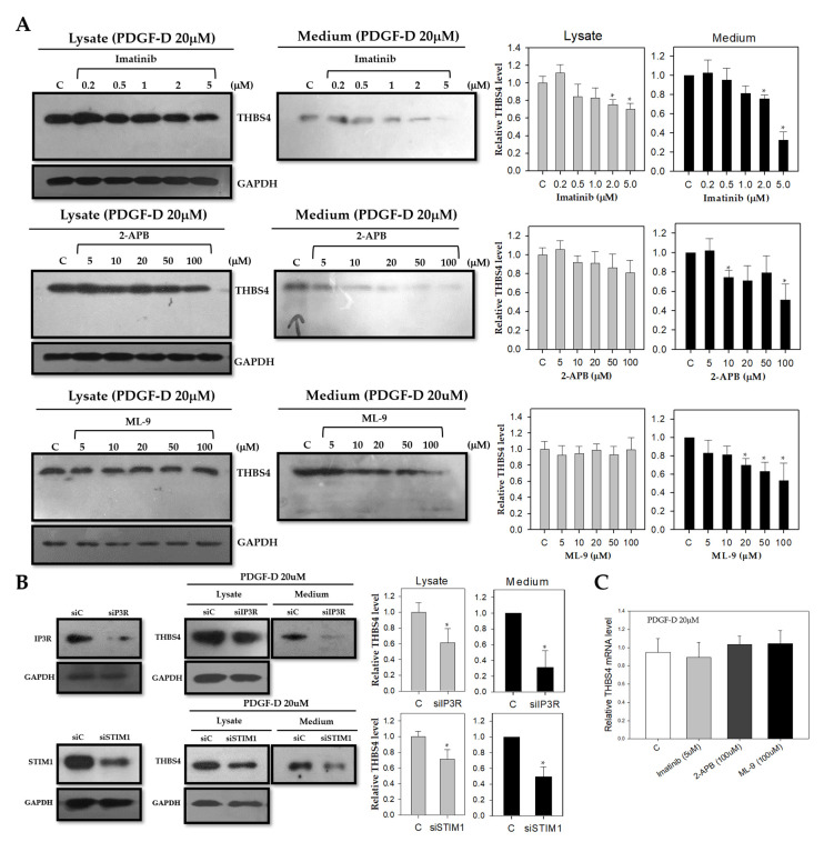
Figure 4.
Effect of PDGF-D stimulation of THBS4 after blockage of PDGFRβ, IP3R, and STIM1. (A) Western blot with anti-THBS4 antibody in whole cell lysate (left panel) or cultured medium (right panel) of DLD-1 cells cultured with PDGF-D (20 μM) for 8 h in the presence of imatinib (0, 0.2, 0.5, 1, 2, and 5 μM) for 16 h, 2-APB (0, 5, 10, 20, 50, and 100 μM) for 16 h, or ML-9 (0, 5, 10, 20, 50, and 100 μM) for 16 h, respectively. (B) Western blot with anti-THBS4 antibody in whole cell lysate or cultured medium of DLD-1 cells transfected with siIP3R or siSTIM1 and cultured with PDGF-D (20 μM) for 8 h. (C) Relative mRNA expression levels as determined with real-time PCR for THBS4 of DLD-1 cells cultured with PDGF-D (20 μM) for 8 h in the presence of imatinib (5 μM) for 16 h, 2-APB (100 μM) for 16 h or ML-9 (100 μM) for 16 h, respectively. Three independent experiments were performed in duplicate. * p < 0.05 when compared with the control t-test.

