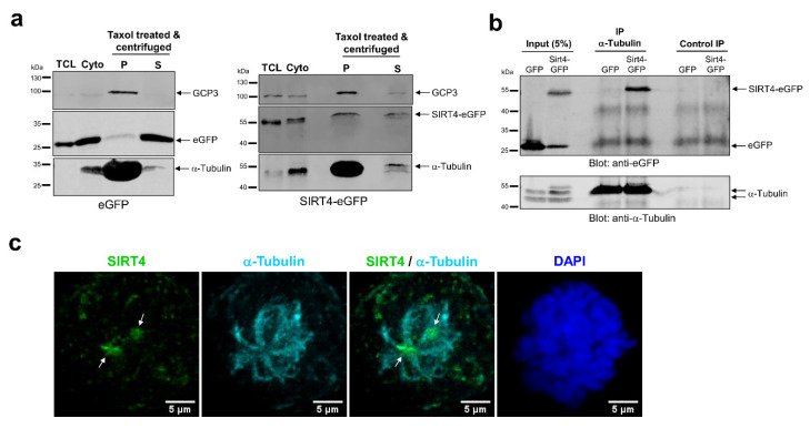
Figure 6.
SIRT4 precipitates with microtubules and co-immunoprecipitates with α-tubulin in HEK293 cells. (a) SIRT4-eGFP, but not eGFP, is present in the pelleted fraction (P) of microtubules which were Taxol-stabilized in the cytosolic fraction (Cyto) followed by pelleting via centrifugation through a sucrose cushion. TCL, Total cell lysate; S, supernatant. Tubulin Gamma Complex Associated Protein 3 (TUBGCP3 or GCP3) was detected as co-marker for microtubules (b). An α-tubulin specific antibody co-immunoprecipitates SIRT4-eGFP, but not eGFP, from total cell lysates of stably transfected HEK293 cells. As control, immunoprecipitation without α-tubulin antibody was performed. (c) Localization of SIRT4 at spindle poles/Microtubule Organizing Centers (MTOCs) of mitotic HeLa cells using a polyclonal antibody against SIRT4 (sc-135053, Santa Cruz Biotechnology, Heidelberg, Germany) and analysis by spinning disk microscopy. Antibodies against α-Tubulin were employed to visualize microtubules. DAPI was used to detect DNA. Bar: 5 µm.

